On a molecular level, muscle contraction and relaxation are achieved when thick filaments containing myosin and thin filaments containing actin interact in the sarcomere—the fundamental functional unit of striated muscle. Myosin-binding protein-C (MyBP-C) is a thick-filament regulatory protein expressed in striated muscle. It has three paralogs, each encoded by a different gene: slow skeletal (sMyBP-C), fast skeletal (fMyBP-C), and cardiac (cMyBP-C). Much effort has been placed toward studying the cardiac isoform due to its role in hypertrophic cardiomyopathy, a disease caused by abnormal genes in the heart muscle, which cause the walls of the heart chamber (left ventricle) to contract harder and become thicker than normal causing the thickened walls to become stiff, while fMyBP-C remains the least studied. However, interest in fMyBP-C is increasing because the fast skeletal isoform has been linked to congenital skeletal muscle diseases. These myopathies include distal arthrogryposis and lethal congenital contracture syndrome, which can cause devastating impairments such as muscle weakness and atrophy and can even be incompatible with life. Recently, a group of researchers set out to better understand fMyBP-C on a molecular level. Using knockout mice, the researchers conducted a suite of experiments including x-ray diffraction, which was carried out at the U.S. Department of Energy’s Advanced Photon Source (APS). Insights from these experiments helped the team elucidate the structural and functional roles that fMyBP-C plays in fast skeletal muscle contraction. These findings, published in the Proceedings of the National Academy of Sciences of the United States of America, elevate the field’s understanding of myosin-binding proteins and move forward research related to skeletal myopathies.
Human skeletal and cardiac muscle is made up of thousands of organelles called myofibrils that allow muscles to contract. As they contract, sliding occurs between the myosin-based thick and actin-based thin filament contractile proteins, allowing the muscles to shorten. Myosin-binding protein-C (MyBP-C) governs this process. The three MyBP-C paralogs, sMyBP-C, fMyBP-C, and cMyBP-C have similar protein structures but are each encoded by different genes and have unique functional and regulatory mechanisms.
To better understand the structural, functional, and physiological roles of fMyBP-C in skeletal muscle, the researchers in this study conducted in vitro and in vivo studies on newly generated homozygous fMyBP-C knockout mice (C2−/−). The C2−/− mice showed decreased grip strength and plantar flexor muscle strength and reduced peak isometric tetanic force and isotonic speed of contraction in isolated extensor digitorum longus (EDL) muscle.
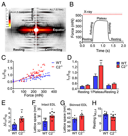
Using the Bio-CAT 18-ID beamline at the APS (the APS is an Office of Science user facility at Argonne National Laboratory) for small-angle x-ray diffraction studies, the researchers measured the x-ray diffraction pattern of EDL muscle both at rest and during isometric contraction. The resulting data (Fig. 1) on the C2−/− EDL muscle showed increased mobility of myosin heads. During contraction, there was a shift of myosin heads toward actin and, at rest, the myosin heads were less ordered. The data also suggested increased spacing in the interfilament lattice spacing for C2−/− EDL muscle.
These findings were corroborated by experiments which examined fMyBP-C myofilament calcium sensitivity. They found that, during Ca2+-activation, C2−/− mice EDL muscle fibers demonstrated decreased steady-state isometric force. They also observed decreased myofilament calcium sensitivity and sinusoidal stiffness. The study also showed that fMyBP-C null muscles respond weakly to mechanical overload, displaying muscle damage and disrupted inflammatory and regenerative pathways.
This study improved our understanding about how the three fMyBP-C paralogs differ in structure and function. Results from this work reveal important functional information about fast-skeletal MyBP-C in fast-twitch muscle: that fMyBP-C regulates force, power, and contractile speed. By regulating myofilament calcium sensitivity and actin–myosin interactions, fMyBP-C produces forces that meet the demands of fast-twitch skeletal muscles. These new insights increase the field’s knowledge of myosin-binding proteins and will accelerate skeletal muscle disease research. ― Alicia Surrao
See: Taejeong Song1, James W. McNamara1‡,‡‡, Weikang Ma2, Maicon Landim-Vieira3, Kyoung Hwan Lee4‡‡‡, Lisa A. Martin1, Judith A. Heiny5, John N. Lorenz5, Roger Craig4, Jose Renato Pinto3, Thomas Irving2, and Sakthivel Sadayappan1*, “Fast skeletal myosin-binding protein-C regulates fast skeletal muscle contraction,” Proc. Natl. Acad. Sci. U.S.A 118(17), e2003596118 (2021). DOI: 10.1073/pnas.2003596118
Author affiliations: 1University of Cincinnati, 2Illinois Institute of Technology, 3Florida State University College of Medicine, 4University of Massachusetts Medical School, 5University of Cincinnati College of Medicine ‡Present addresses: The Royal Children’s Hospital, ‡‡The University of Melbourne, ‡‡‡ University of Massachusetts Medical School
Correspondence: * sadayasl@ucmail.uc.edu
S.S. has received support from National Institutes of Health (NIH) grants R01 HL130356, R01 HL105826, R01 AR078001, and R01 HL143490; and American Heart Association (AHA) 2019 Institutional Undergraduate Student (19UFEL34380251) and transformation (19TPA34830084) awards. T.S. (19POST34380448) and J.W.M. (17POST33630095) were supported with AHA Fellowship training grants. R.C. was supported by NIH grants P01 HL059408, R01 AR067279, and R01 HL139883. Bio-CAT is supported by grant P41 GM103622 from the National Institute of General Medical Sciences of the NIH. Through Loyola University Chicago, Pieter de Tombe provided institutional funding support to generate C2−/− mice. This research used resources of the Advanced Photon Source, a U.S. DOE Office of Science User Facility operated for the DOE Office of Science by Argonne National Laboratory under contract no. DE-AC02-06CH11357.
The U.S. Department of Energy's APS is one of the world’s most productive x-ray light source facilities. Each year, the APS provides high-brightness x-ray beams to a diverse community of more than 5,000 researchers in materials science, chemistry, condensed matter physics, the life and environmental sciences, and applied research. Researchers using the APS produce over 2,000 publications each year detailing impactful discoveries, and solve more vital biological protein structures than users of any other x-ray light source research facility. APS x-rays are ideally suited for explorations of materials and biological structures; elemental distribution; chemical, magnetic, electronic states; and a wide range of technologically important engineering systems from batteries to fuel injector sprays, all of which are the foundations of our nation’s economic, technological, and physical well-being.
Argonne National Laboratory seeks solutions to pressing national problems in science and technology. The nation's first national laboratory, Argonne conducts leading-edge basic and applied scientific research in virtually every scientific discipline. Argonne researchers work closely with researchers from hundreds of companies, universities, and federal, state and municipal agencies to help them solve their specific problems, advance America's scientific leadership and prepare the nation for a better future. With employees from more than 60 nations, Argonne is managed by UChicago Argonne, LLC, for the U.S. DOE Office of Science.
The U.S. Department of Energy's Office of Science is the single largest supporter of basic research in the physical sciences in the United States and is working to address some of the most pressing challenges of our time. For more information, visit the Office of Science website.
