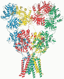
The full structure of a fiendishly complicated and important brain protein has been determined by researchers using two U.S. Department of Energy (DOE) light sources, potentially enabling the development of new treatments for a wealth of neurological disorders.
Eric Gouaux and his colleagues undertook the difficult task of mapping the structure of a glutamate receptor, a protein that mediates signaling between neurons in the brain and elsewhere in the nervous system. The receptor is also thought to be crucial to processes such as memory and learning.
The resulting picture "tells us things about the organization of the receptor that were just completely unanticipated" says Gouaux, a protein crystallographer at Oregon Health and Science University in Portland. The work appears online today in Nature1.
The researchers studied a rat glutamate receptor known as GluA2. They grew a crystal of many such proteins, then exposed this to a beam of x-rays. By watching how x-rays scattered from the crystal, they were able to produce an atomic-level picture of a single protein. [X-ray diffraction data were obtained at Northeastern Collaborative Access Team beamline 24-ID-E of the Advanced Photon Source at the DOE’s Argonne National Laboratory and beamlines 5.0.2, 8.2.1, and 8.2.2 of the Advanced Light Source at the DOE’s Lawrence Berkeley National Laboratory.]
In humans, these receptors work as relays for the central nervous system. When the neurotransmitter glutamate binds to the receptor, this opens an 'ion channel' in the neuronal membrane, allowing ions to flow across the membrane. This results in the transmission of an electrical pulse down the nerve.
Until now, the structure of an intact glutamate receptor has never been seen. Knowing its shape will not only allow scientists to better understand how it works, but should also help those working to develop therapies for conditions in which something goes awry with the system, such as epilepsy and Alzheimer's disease.
The shape of this receptor can now be taken into account when designing molecules that could function as drugs by binding to it. "If you know what the lock looks like then you can better design a key," says Gouaux. "If you don't know you're at a loss."
The GluA2 receptor is shaped like a capital letter Y, and has three main parts. At the top are two prongs, which can enable modification of the receptor. Below this is the area where glutamate binds, triggering the opening of the ion channel. And at the bottom is the channel itself, shaped, say the authors, "like a Mayan temple."
The receptor comprises four subunits, which are chemically identical but, surprisingly, are folded differently (see picture). "The completely astonishing thing was that two subunits are completely different from the other two," says Gouaux. "That difference was totally unanticipated."
Stephen Traynelis, head of a lab that specializes in glutamate receptors at Emory University in Atlanta, Georgia, says that the structure will be a huge boon to research into receptor biology. Previous work involved making inferences — now that researchers have a complete receptor mapped, they can go back and reinterpret their previous research in light of this.
"This will be enormously useful," Traynelis says. "It's hard to overstate its importance. It's probably one of the most important papers that have come out in this field in the last 10 years."
Gouaux's paper presents the structure with the channel closed. Traynelis says a key future goal is to analyse the structure of the receptor when the ion channel is open.
Crystallizing any membrane protein to allow its structure to be determined with X-rays is a major challenge, says Elek Molnar, a neuroscientist who works on glutamate receptors at the University of Bristol, UK. In the case of this "excellent paper," the researchers chemically locked the receptor into one conformation, with the ion channel closed, to assist in crystallization and visualization.
Future work should allow researchers to see how the receptor interacts with other elements of the nervous system, such as auxiliary proteins. "It is kind of like providing a skeleton," says Molnar. "Now future research can attach muscles, tendons and so on."
Reprinted by permission from Macmillan Publishers Ltd.: “naturenews.” Published online 29 November 2009 | Nature | doi:10.1038/news.2009.1114 Copyright 2009.
See: Alexander I. Sobolevsky1, Michael P. Rosconi1,2, and Eric Gouaux1*, “X-ray structure, symmetry and mechanism of an AMPA-subtype glutamate receptor,” Nature advance online publication 29 November 2009 | doi:10.1038.
Author affiliations: 1Oregon Health and Science University, 2Present address: Regeneron Pharmaceuticals, Inc.
Correspondence: gouauxe@ohsu.edu).
M.P.R. was supported by an individual NIH National Research Service Award. This work was supported by the NIH. E.G. is an investigator with the Howard Hughes Medical Institute. Use of the Advanced Photon Source at Argonne National Laboratory was supported by the U. S. Department of Energy, Office of Science, Office of Basic Energy Sciences, under Contract No. DE-AC02-06CH11357.

