
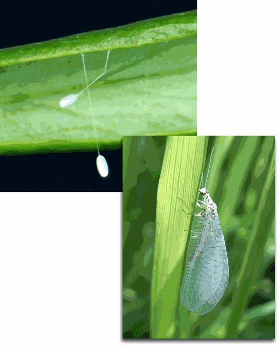
Green lacewings (Chrysopidae) are beneficial insects well known to naturalists and scientists for their many elegant solutions to life’s problems, such as how to eat without being eaten. The larvae of many species adopt a wolf-in-sheep’s-clothing strategy when feeding upon their major prey item, aphids. They cover themselves with either the tufted wax exudate or the spent carcasses of their prey in order to escape detection by honeydew-feeding ants, which normally attack intruders in their aphid herds. The adult female protects her eggs from detection and predation (especially by ants) by suspending each egg at the end of a fine strand of silk (top figure). Utilizing the high-flux ChemMatCARS 15-ID beamline at the APS, researchers from Australia's Commonwealth Scientific and Industrial Research Organisation (CSIRO) recently completed studies that confirm the elegant and unique structure of the lacewing’s silken egg stalk. The information they obtained suggests that lacewing silk may have potential value as a biomaterial.
“Lacewing egg stalk silk is based on completely different design principles to the well-known silks of domesticated silkworms or spider webs,” said lead researcher Sarah Weisman. “The lacewing silk evolved to function as a rigid support column, rather than a long flexible thread. Its protein sequence and structure are unique.”
The scientists studied Mallada signata, a species of green lacewing native to Australia. A half-century earlier, classic protein structural studies had reported on the unusual cross-beta sheet structure of the silk of another green lacewing species, Chrysopa flava. The CSIRO researchers were able to apply the technological advances of recent decades to identifying the genes that encode the silk proteins and developing a more detailed model of the structure.
“We used wide-angle x-ray scattering at the ChemMatCARS facility to give us detailed information about the protein structure of a single silk fiber,” Weisman said. “Since the tiny egg stalks are only 3 millimeters long and 15 microns in diameter, we couldn't have obtained clear data anywhere except at a high-flux third-generation synchrotron source.”
Functional proteins with a cross-beta structure are rare in nature and appear to be limited to extruded products such as arthropod silks. Within a cellular environment, cross-beta structures occur as errors in protein folding. These misfolded proteins can aggregate to form the amyloid plaques that characterize neurodegenerative diseases such as Alzheimer’s and Parkinson’s. Accordingly, cross-beta structures are of great interest to researchers in diverse fields.
The CSIRO researchers identified and sequenced two silk genes, which they named MalXB1 and MalXB2. These genes are expressed in adult female M signata only, not in males or larvae, confirming that the silk of lacewing cocoons is composed of different proteins. Over 70% of each protein is composed of repeating 16-residue motifs, suggesting a high level of crystallinity. Based on their data, the CSIRO researchers propose that the cross-beta structure is made up of 8-residue crystallite strands stacked perpendicular to the direction of the fiber. As the lacewing female draws the liquid silk into a strand, a network of cysteine cross links are created that result in rapid hardening of the stalk.
The cross-beta structure accounts for the remarkable extensibility of the egg stalk silk, which can be stretched to nearly 500% of its length before breaking. Its bending rigidity, achieved by cysteine cross linking, is almost three times greater than that of silkworm fibers. These properties, along with reasonable tensile strength and the relative simplicity of its production, suggest lacewing silk may have potential value as a biomaterial. — Carol Hart
See: Sarah Weisman1, Shoko Okada1, Stephen T. Mudie2, Mickey G. Huson2, Holly E. Trueman1, Alagacone Sriskantha1, Victoria S. Haritos1, and Tara D. Sutherland1*, “Fifty Years Later: The Sequence, Structure and Function of Lacewing Cross-Beta Silk,” J. Struct. Biol. 168, 467 (2009).
DOI: 10.1016/j.jsb.2009.07.002
Author Affiliations: 1CSIRO Entomology, 2CSIRO Material Science and Engineering
Correspondence: *Tara.Sutherland@csiro.au
This work was financially supported by the Grains Research and Development Corporation. Use of ChemMatCARS Sector 15 at the Advanced Photon Source was supported by the Australian Synchrotron Research Program, which is funded by the Commonwealth of Australia under the Major National Research Facilities Program. ChemMatCARS Sector 15 is principally supported by the National Science Foundation/Department of Energy under Grant No. CHE-0535644.
Use of the Advanced Photon Source is supported by the U.S. Department of Energy, Office of Science, Office of Basic Energy Sciences, under Contract No. DE-AC02-06CH11357.
Argonne National Laboratory seeks solutions to pressing national problems in science and technology. The nation's first national laboratory, Argonne conducts leading-edge basic and applied scientific research in virtually every scientific discipline. Argonne researchers work closely with researchers from hundreds of companies, universities, and federal, state and municipal agencies to help them solve their specific problems, advance America 's scientific leadership and prepare the nation for a better future. With employees from more than 60 nations, Argonne is managed by UChicago Argonne, LLC for the U.S. Department of Energy's Office of Science.