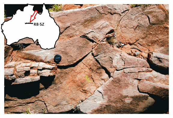
A team of geologists and geophysicists using the U.S. Department of Energy’s Advanced Photon Source (APS) at Argonne National Laboratory has shed light on a fluid transfer in the middle continental crust, a phenomenon that was formerly poorly understood. Their work could lead to a fuller understanding of fluid migration through rock, with possible wide application in deciphering important geological processes including earthquakes.
The movement and behavior of fluid—generally water, sometimes other types—is a prime factor in geological processes including earthquake nucleation, mantle degassing, and the formation of mineral deposits. The workings of fluid transfer in Earth’s middle crust have been controversial and not well understood, although it has been clear that they are associated with shear zones where rock deforms viscously. When it comes to fluid migration, the middle crust is different from other crustal regions because it is generally thought to be both too hot and too viscous to allow fluid to pass through fractures.
The researchers from the University of Western Australia, CSIRO Exploration & Mining, and Argonne National Laboratory have confirmed that a phenomenon called “creep cavitation” is instrumental in the deformation of rock and the creation of porosity that allows fluid to permeate into these mid-crustal shear zones, ultimately leading to large-scale structural weaknesses that can cause fault slippage, and even slow earthquakes. The team has developed a model for a “granular fluid pump” driven by stress gradients and chemical potentials on the grain scale that can move fluid through rocks deforming at mid-crustal conditions.
The research team used synchrotron x-ray microtomography at the X-ray Operations and Research 2-BM x-ray beamline to study a characteristic rock sample collected from the Redbank midcrustal shear zone in Central Australia. Examining 55-mm-sized rock cubes culled from various points of a rock hand specimen, the team was able to visualize the structure of all pores larger than 1.3-µm in diameter. They could thus obtain a detailed picture of the porosity architecture of the entire rock sample.
The rock sample covered a strain gradient, allowing study and comparison of the evolution of porosity from the low- to high-strain regions. This strain gradient was traced by the microstructural transformation of the rock from a gneiss to a mylonite: low-strain regions are largely characterized by grains of K-feldspar and plagioclase minerals (hundreds of microns in diameter) interlayered with quartz bands, while towards the more deformed areas, this microfabric of the rock gives way to a finer-grained (>10 µm) homogenized mylonite pattern. The reduction in grain size in the high-strain areas of the sample shows that this rock was progressively softened as it deformed, and the gradual disappearance of K-feldspar and plagioclase indicates that they were dissolved and their chemical components redistributed through the rock by a fluid at the same time.
For the researchers, this realization pointed the way to their “granular fluid pump” model. Micron-scale grains in the high-strain regions of the rock slide past each other under stress, in a process called viscous grain-boundary sliding (VGBS). As this happens, pores open up while others close in the rock structure, to accommodate incompatibilities that arise between neighboring grains. This mechanism is called creep cavitation, and has been observed in some ceramic and metallic materials. In the middle crust, fluid moves from the closing to the opening pores as the fluid pressure changes from pore to pore during creep cavitation. This mechanism is supported by chemical reactions between the fluid and the minerals forming the rock; as minerals get dissolved in, or precipitated from the fluid, the fluid pressure changes locally. The resulting granular fluid pump is self-sustaining and steady as long as grains slide past each other, and creep cavitation occurs. The experimenters speculate that aside from affecting rock deformation on the small scale, the same combination of processes—creep cavitation and the granular fluid pump could—on a greater scale, influence phenomena such as fault slippage and slow earthquakes.
The work represents the first time that x-ray synchrotron tomography has been used to visualize porosity in a mid-crustal shear zone rock at such small scale. “None of us had previously used tomography to visualize micron-sized pores in crystalline rocks,” said lead author Florian Fusseis. “We were quite blown away how our bravest expectations were met by the results.” The x-ray tomography allowed the researchers to clearly follow the evolution of the porosity of the sample during rock deformation and quantify it with great precision. “Synchrotron tomography is obviously the perfect technique to investigate porosity in crystalline rocks,” said Fusseis.
Using this technique, the researchers have managed to shed light on a phenomenon that was formerly poorly understood and to develop a model with possible wide application. “I believe that our granular fluid pump applies to many tectonic settings and explains fluid transfer in specific rock types quite well, and is as such quite significant,” Fusseis said.
Fusseis and his team plan to expand upon the work by examining further mid-crustal shear zone samples at even finer resolutions of approximately 35 nm at the APS Sector 32 beamline. Along with the collection of this more detailed data, and comparison and study of different samples from other parts of the world, the researchers hope to use the micro- and nanotomography capabilities of the APS to observe porosity formation in real-time controlled experiments. This multipronged approach will lead to a better understanding of the highly complex and sometimes mysterious workings of the Earth’s middle crustal zones. — Mark Wolverton
See: F. Fusseis1,3*, K. Regenauer-Lieb1,2,3, J. Liu2, R.M. Hough2, and F. De Carlo4, "Creep cavitation can establish a dynamic granular fluid pump in ductile shear zones," Nature 459, 975 (18 June, 2009). DOI: 10.1038/nature08051
Author affiliations: 1 The University of Western Australia, 2CSIRO Exploration & Mining, 3Western Australian Geothermal Centre of Excellence, 4Argonne National Laboratory
Correspondence: *fusseis@cyllene.uwa.edu.au
This work was supported by the Australian Synchrotron Research Program, which is funded by the Commonwealth of Australia under the Major National Research Facilities Program; the Western Australian Premier’s Research Fellowship program and the University of Western Australia through a research grant; iVEC through the use of visualization resources and expertise provided by the WASP and ARRC facilities; and the Centre for Microscopy, Characterization and Analysis at the University of Western Australia for the use of its FESEM. Use of the Advanced Photon Source at Argonne National Laboratory was supported by the US Department of Energy, Office of Science, Office of Basic Energy Sciences, under contract number DE-AC02-06CH11357.

